Why Our Baby? TRISOMY 13/18 CHROMOSOMES
This chapter sets out to outline the science of chromosomes and give the reader some information on chromosomes and how they are relevant for our Edwards’ T18 and Patau T13 babies.
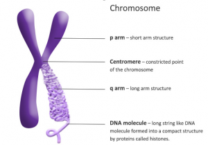
Chromosomes are tiny structures inside cells made from DNA and protein. The information inside chromosomes acts like a recipe that tells cells how to function and replicate. Every form of life has its own unique set of instructions. Each chromosome consists of two parts: the short arm “p” and the long arm “q”. On the chromosomes there are between 50,000 and 100,000 genes. These genes determine the individual hereditary characteristics of each person i.e., height, eye colour, blood group. See Fig 1
A human cell contains 46 chromosomes and these are arranged into 23 pairs. A human egg or sperm contains only one chromosome from each pair. Therefore, there should be 23 of the mother’s chromosomes in each egg and 23 of the father’s chromosomes in each sperm, the resultant fertilised egg will contain 46 chromosomes (23 pairs); one chromosome of each pair came from the mother and the other chromosome from the father. This is the unique “blueprint” for the individual baby. See Fig 2
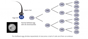
Figure 2. The fertilised egg divides repeatedly to become first a ball of cells and then an embryo.
Karyotypes
A karyotype is an individual’s collection of chromosomes. The term also refers to a laboratory technique that produces an image of an individual’s chromosomes. The karyotype is used to look for abnormal numbers or structures of chromosomes. Figure 3 below shows the structure of a male with the correct number of chromosomes.
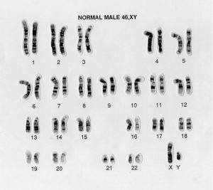
Figure 3. This is a karyotype of a male with the correct number of chromosomes.
Trisomy – Definition and occurrence
Trisomy is a condition in which an extra copy of a chromosome is present in the cell nuclei, causing developmental abnormalities.
In Trisomy 13 (Patau’s syndrome) an extra chromosome number 13 is present in each cell. See Fig 4
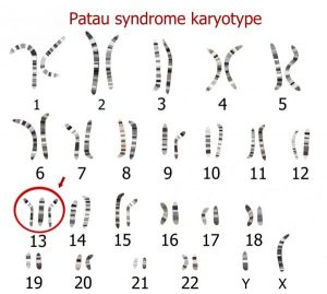
Figure 4. The karyotype of a male with Trisomy 13.
In Trisomy 18 (Edwards’ syndrome) an extra chromosome number 18 is present in each cell. See Fig 5
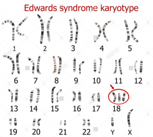
Figure 5. The karyotype of a female with Trisomy 18.
The extra chromosome may come from either the mother’s egg or from the father’s sperm during cell formation, or when the cells of the fertilised egg do not divide correctly during cell division. See Fig 6
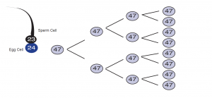
Figure 6. Here you can see that the chromosomes did not divide correctly. The result is an extra chromosome or trisomy. Trisomy may occur in either the mother’s egg or the father’s sperm.
Although the genes in the three chromosomes are normal, too much or too little genetic material in a cell affects every stage of the development of the baby. Therefore the blueprint for development is altered from the moment of conception.
Related Disorders
Both Trisomy 13 and Trisomy 18, have a number of linked disorders that can be associated with them. These are outlined below
Partial Trisomy
A very small percentage of babies are born, not with a complete trisomy, but with a partial trisomy. This is much rarer and means that the affected baby has TWO copies of the affected chromosome PLUS a partial copy. Babies with a partial trisomy may show some or all of the characteristics of the particular trisomy. This depends on the precise amount and nature of the additional chromosomal material present.
Why Do Translocations Happen?
Although about 1 person in 500 has a translocation, it is still not really understood why they happen. It is known that chromosomes seem to break and re-join quite often during the making of sperm and eggs or around the time of conception, and it is only sometimes that this leads to problems. These changes occur without us being able to control them.
Balanced Translocation
In a balanced translocation, the normal number (46) of chromosomes are present in each cell but some or all of the material from one chromosome may be located onto a different chromosome. A person who carries a balanced translocation is not usually affected by it, and is often unaware of having it. There is no loss of genetic material. Babies with balance translocations are completely normal and their chromosomal rearrangement should have no implications regarding health or physical appearance. The only time it may become important is if he or she has children. This is because the child may inherit what we call an unbalanced translocation
Unbalanced Translocation
This means that either
(a) an extra piece of chromosomal material is present in each cell, usually attached to another chromosome
(b) that a piece of chromosomal material is missing.
Balanced or unbalanced translocations can arise spontaneously where both parents have normal chromosomes. However, if a parent has a balanced chromosome re-arrangement then there can be a higher chance of that person having a pregnancy that may result in a miscarriage or also going on to have a pregnancy that is affected with an unbalanced chromosome translocation. The chance of these happening depends on the nature of the translocation and what has previously happened in the family.
In addition, where one person in the family carries a balanced translocation it is also possible that other members of the family, who are otherwise healthy, may also carry that translocation, which may have implications in turn for them if they are thinking about having children.
Deletion
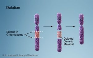
A deletion is a condition where some chromosomal material is missing from the normal chromosome. In the case of a condition denoted by 13q-, the long arm “q” of chromosome 13 is missing. In the case of 18p- the short arm “p” of chromosome 18 is missing. See Fig. 7
The severity of the condition and the signs and symptoms depend on the size and location of the deletion and which genes are involved.
Figure 7
Mosaicism
This condition is a less common form of trisomy. It results from an incorrect separation of chromosomes after the first normal division of the fertilised egg. Therefore, some cells have an extra chromosome (47), while other cells have the normal number of chromosomes (46). The number of cells having the extra chromosome varies from baby to baby and therefore each baby with mosaicism is unique. These babies are often less severely affected compared to those with an extra chromosome in every cell
Ring Formation
Ring Chromosome is a rare chromosome abnormality in which the ends (arms) of the chromosome join together to form a ring shape.
When a ring chromosome forms, genetic material can be lost from either arm or both arms, causing various signs and symptoms. People with ring chromosome mosaicism may have milder symptoms. See Figure 8
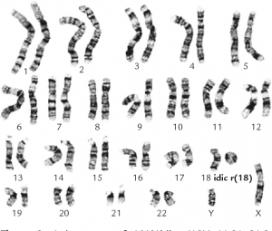
Figure 8
Genetic counselling
A genetic counselling service is available to all parents but there are waiting lists. Your GP, obstetrician, or paediatrician can refer you for counselling. Your GP may refer you for genetic counselling, if appropriate, to the Department of Clinical Genetics, Our Lady’s Children’s Hospital, Crumlin, Dublin 12.
The genetic counsellor will:
- Discuss the results of your baby’s chromosome study.
- Explain all aspects of the disorder.
- Discuss the variable and less common forms of trisomy with the family.
- Assess the risk of having a second baby with a chromosomal disorder.
- Discuss the implications of the genetic diagnosis for any children or any other family members that the couple may have.
- Answer any questions you may have.
In most cases, the genetic counsellor will be able to reassure you that Patau’s or Edwards’ syndrome is unlikely to reoccur.
